Penetrating Neck Injury
Trauma     All Diseases     eMedicine
General Information:
Penetrating neck injury: Trauma involving a missile or sharp object penetrating the skin and violating the platysma layer of the neck.

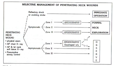
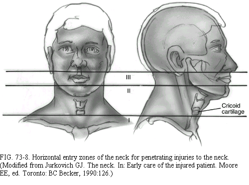
Management has changed significantly over the years The management now focuses on the zones of the neck (see above) and the stability of the patient.
HPI/Physical
Significant injuries needing OR:
•    Air bubbling through the wound
•    Active pulsatile bleeding
•    Expanding Hematoma
•    Neurologic deficit
•    Hypotension/Unstable vitals
Signs of airway injury:
•    Subcutaneous air
•    Air bubbling through the wound
•    Stridor or respiratory distress
Signs of vascular injury:
•    Expanding Hematoma
•    Active pulsatile bleeding
•    Bruit/thrill
•    Pulse deficit
Management
Initial management:
ATLS resuscitation,  IV Access, intubation if indicated
Circulation, Airway, Breathing.
Looking for injury to airway.
C-spine immobilization is contraindicated

-Possible benefit for lowering BP to MAP of 50 mm of Hg to help with hemorrhage control.

Aggressive resuscitation may elevate the blood pressure and increase hemorrhaging of the injury site.

All patients get portable Chest X-ray and AP & Lat Neck
Unstable Patient:  
Immediate surgical exploration in the OR for patients who present with signs and symptoms of shock and continuous hemorrhage from the neck wound.
Surgical management varies by zone; zone I and zone III are difficult to expose making vascular control problematic, leading to higher mortality in zone I and III.
Stable Patient:
The laceration should never be probed or locally explored in the emergency department if platysma is violated.
This could dislodge a clot and initiate hemorrhage.
If no significant injuries requiring surgery are present, surgery is not indicated and the patient should be observed.


Zone I Neck Injury Zone II Neck Injury Zone III Neck Injury
Stable & Symptomatic: Four-vessel cerebral angiography is indicated and OR for surgical exploration
Angiography remains the standard for assessing for arterial anatomy and injury
- CTA possible alternative in these patients

Stable & Asymptomatic:
Angiography, esophagiscopy  or esophagram
- CT considered as replacement (2)
Stable & Symptomatic: OR for exploration

Stable & Asymptomatic: Observe
CT evaluation considered as adjunct (2)
Stable & Symptomatic: Four-vessel cerebral angiography is indicated and OR for exploration
Angiography remains the standard for assessing for arterial anatomy and injury
- CTA possible alternative in these patients (1)
Stable & Asymptomatic: Observation
CT considered as adjunct (2)
Ferguson et al. suggested angiography is not necessary if no hard vascular signs in zone III(3)